Medical Papers Cervical

Option for Transverse Midline Incision and Other Factors That Determine Patient’s Decision to Have Cervical Spine Surgery

By Dr. Kingsley Chin

LI_BlogCover_CervicalTransverseIncision_v05-700x400.jpg
“I had my [cervical fusion and lumbar decompression] surgery ten days ago and I feel great, I’m wearing my heels and I’m ready to go!”

Introductory content by Fabio Pencle 

“[Dr. Chin] is a true professional… the whole team, the whole staff was great here at the LESS Institute.”

“[Dr. Chin] is a true professional… the whole team, the whole staff was great here at the LESS Institute.”

A newly published study has demonstrated that patients prefer a midline incision for cervical spine surgery.

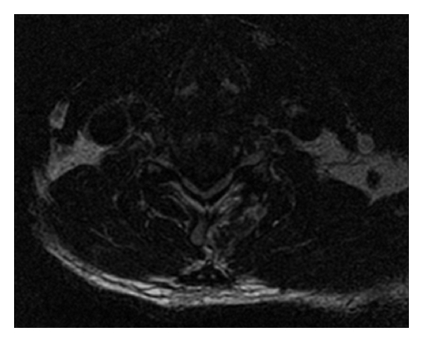
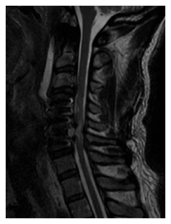
The anterior approach to cervical spine surgery has become the standard for the treatment for degenerative disc, traumatic herniated disc and fracture dislocation. Cloward, Smith and Robinson have

devised operative techniques with modifications by several surgeons since 1958. There are varying types of skin incisions for cervical spine surgery; the incision is either on the right or left side of the trachea based on the surgical approach to the recurrent laryngeal nerve. Other factors determining the type of incision include a few pathological levels affected, if corpectomy is required and whetheraffected segments are contiguous.

Transverse midline incisions have been used by other surgical specialties such as ENT, vascular and general surgeons. This incision provides a more cosmetically acceptable result and allows for access to structures during surgery; however, few studies discuss the relevance of cosmesis. There are several named guidelines for determining surgical incision, most notably, Langer’s lines. A transverse midline incision would, however, follow the guideline by Kraissl, where the incision is made in a skin crease. The quality of surgery is judged immediately by the amount of relief of symptoms and the cosmetic.

Considering the patient-driven procedures offered to treat the same pathology, as well as recent trends in the increase in ambulatory surgery center (ASC) use, the authors felt it prudent to devise a questionnaire with the primary goal of determining the preferences of the patients. The secondary goal was to determine factors which lead to the decision to have anterior cervical spine surgery.

Scientific Paper

Fabio J.R. Pencle, Jason A. Seale, Amala Benny, Sephania Salomon, Ashley Simela, Kingsley R. Chin

To read the full paper & citations as published in the Journal of Orthopaedics, visit here.

Background

Authors aim to determine patients’ preference for surgical incision and factors affecting the decision for surgery to the anterior neck.

Methods

A questionnaire was presented prior to evaluation and if preceded to surgery followup given.

Results

243 patients completed questionnaire, with 60% female population and younger than 50 years. 151 patients preferred a transverse midline incision with a statistically significant increase in outcomes and cosmesis importance and a decrease in the importance of board certification.

Conclusion

Findings of questionnaire demonstrate that patients’ prefer a transverse midline anterior neck incision, with surgical outcomes being the overall factor affecting decision making.

About Author Dr. Kingsley R. Chin

Dr. Kingsley R. Chin, Founder of philosophy and practice of The LES Society and The LESS Institute

Dr. Kingsley R. Chin, Founder of philosophy and practice of The LES Society and The LESS Institute

Dr. Kingsley R. Chin is a board certified Harvard-trained orthopedic spine surgeon and professor with copious business and information technology exposure. He sees a niche opportunity where medicine, business and info. tech meet – and is uniquely educated at the intersection of these three professions. He has experience as Professor of Clinical Biomedical Sciences & Admissions Committee Member at the Charles E. Schmidt College of Medicine at Florida Atlantic University, Professor of Clinical Orthopedic Surgery at the Herbert Wertheim College of Medicine at Florida International University, Assistant Professor of Orthopaedics at the University of Pennsylvania Medical School, VisitingSpine Surgeon & Professor at the University of the West Indies, Mona, and Adjunct Professor of Clinical Biomedical Studies at the University of Technology, Jamaica.

Learn more about Dr. Chin here and connect via LinkedIn.

About Less Exposure Surgery

Less Exposure Surgery (LES) is based on a new philosophy of performing surgery, leading the charge to prove through bench and clinical outcomes research that LES treatment options are the best solutions – to lowering the cost of healthcare, improving outcomes and increasing patient satisfaction. Learn more at LESSociety.org.

The LES Society philosophy: “Tailor treatment to the individual aiding in the quickest recovery and return to a pain-free lifestyle, using LES® techniques that lessen exposure, preserve unoffending anatomy and utilize new technologies which are safe, easy to adopt and reproducible. These LES®techniques lessen blood loss, surgical time and exposure to radiation and can be safely performed in an outpatient center. Less is more.” – Kingsley R. Chin, MD

About The LESS Institute

The LESS Institute is the world leader center of excellence in Less Exposure Surgery. Our safe, effective outpatient treatments help patients recover quickly, avoid expensive hospital stays and return home to their family the same day. Watch our patient stories, follow us on Facebook and visit TheLESSInstitute.com to learn more.

Scientific Paper Author & Citation Details

Authors

Fabio J.R. Penclead, Jason A. Sealead, Amala Bennyd, Sephania Salomond, Ashley Simelade, Kingsley R. Chinabcf

Author information

a. Less Exposure Surgery Specialists Institute (LESS Institute), United States
b. Herbert Wertheim College of Medicine, Florida International University, United States
c. Charles E. Schmidt College of Medicine, Florida Atlantic University, United States
d. Less Exposure Surgery (LES) Society, United States
e. Bronx Lebanon Hospital Center, United States
f. University of Technology, Jamaica


Decreasing Radiation Dose With FluoroLESS Standalone Anterior Cervical Fusion

By Dr. Kingsley Chin

LI_Social_Blog_FlourolessPaper.jpg

Scientific Paper

Kingsley R. Chin, MD, Fabio J.R. Pencle, MB BS, Jason A. Seale, MB BS

Interested medical professionals can read through the full paper, as published in the Journal of Spine Surgery, here.

Study Design

Level III

Objective

Adjacent segment disease and dysphagia remain concerns of anterior cervical discectomy and fusion (ACDF) with fixation using anterior cervical plates (ACPs). The authors aim to demonstrate the feasibility, outcomes and fusion rate of a standalone PEEK cage in the outpatient setting.

Methods

The medical records of 48 consecutive patients undergoing single level standalone ACDF (S-ACDF) (Group 1) were compared to our control group of 49 patients who had single-level ACDF with ACP (Group 2). Outcomes assessed included VAS neck and arm, NDI scores, and radiation dose.

LI_Social_Linkedin_FlourolessPaper_v02.jpg

Results

Forty eight patients in Group 1 (S-ACDF) and 49 patients in Group 2 (ACDF-ACP). No statistical differences in gender, age or BMI were found between groups, p=0.286, 0.691 and 0.947 respectively. There was no intergroup statistically significant difference in preoperative and postoperative outcomes. Mean radiation dose in group 1 of 24.1+/-8.2mAS and 2.0+/-0.7mSv was significantly less compared to group 2 which was 29.8+/-5.4 and 2.5+/-0.5mSv, p<0.001. The average radiation dose for single level fusion in Group 1 was 15.6+/-1.5 mAs and 1.3+/-0.1mSv this is compared to average radiation dose in Group 2 of 27.8+/-3.9mAs and 2.3+/-0.3mSv, p=0.001. The average radiation dose for two level fusion in Group 1 was 30.9+/-3.5 mAs and 2.6+/-0.3mSv this is compared to average radiation dose in Group 2 of 33.9+/-6.0 and 2.9+/-0.5mSV, p=0.012.

Conclusion

In the outpatient setting, S-ACDF has shown statistically significant intergroup difference in overall radiation dose, as well as single and two-level fusions, (p<0.001). We conclude that S-ACDF can decrease overall radiation exposure to patients. This is comparable to single level ACDF-ACP in the outpatient setting.

About Author Dr. Kingsley R. Chin

Dr. Kingsley R. Chin, Founder of philosophy and practice of The LES Society and The LESS Institute

Dr. Kingsley R. Chin, Founder of philosophy and practice of The LES Society and The LESS Institute

Dr. Kingsley R. Chin is a board-certified Harvard-trained Orthopedic Spine Surgeon and Professor with copious business and information technology exposure. He sees a niche opportunity where medicine, business and info. tech meet – and is uniquely educated at the intersection of these three professions. He has experience as Professor of Clinical Biomedical Sciences & Admissions Committee Member at the Charles E. Schmidt College of Medicine at Florida Atlantic University, Professor of Clinical Orthopedic Surgery at the Herbert Wertheim College of Medicine at Florida International University, Assistant Professor of Orthopaedics at the University of Pennsylvania Medical School, Visiting Spine Surgeon & Professor at the University of the West Indies, Mona, and Adjunct Professor of Clinical Biomedical Sciences at the University of Technology, Jamaica.

Learn more about Dr. Chin here and connect via LinkedIn.

About Less Exposure Surgery

Less Exposure Surgery (LES) is based on a new philosophy of performing surgery, leading the charge to prove through bench and clinical outcomes research that LES treatment options are the best solutions – to lowering the cost of healthcare, improving outcomes and increasing patient satisfaction. Learn more at LESSociety.org.

The LES Society philosophy: “Tailor treatment to the individual aiding in the quickest recovery and return to a pain-free lifestyle, using LES® techniques that lessen exposure, preserve unoffending anatomy and utilize new technologies which are safe, easy to adopt and reproducible. These LES®techniques lessen blood loss, surgical time and exposure to radiation and can be safely performed in an outpatient center. Less is more.” – Kingsley R. Chin, MD

About The LESS Institute

The LESS Institute is the world leader center of excellence in Less Exposure Surgery. Our safe, effective outpatient treatments help patients recover quickly, avoid expensive hospital stays and return home to their family the same day. Watch our patient stories, follow us on Facebook and visit TheLESSInstitute.com to learn more.

Scientific Paper Author & Citation Details

Authors

Kingsley R. Chin, MD1,2,3,Fabio J.R. Pencle, MB BS3, Jason A. Seale, MB BS3

Author information

1. Herbert Wertheim College of Medicine at Florida International University

2. Charles E. Schmidt College of Medicine at Florida Atlantic University

3. Less Exposure Surgery (LES) Society

4. Less Exposure Surgery Specialists Institute (LESS Institute).

Misaligned Versus Straight Placement of Anterior Cervical Plates: A Clinical and Radiologic Outcomes Study, JAAOS

By Dr. Kingsley Chin

LI_KRCCervicalPlateAlignmentPaper_Comparison-700x436.png

Scientific Paper

Chin KR1, Pencle FJFrancis SDFrancis CASeale JAHothem EA.

Interested medical professionals can read through the full paper, published in the Journal of the AAOS, here.

Background

In anterior cervical diskectomy and fusion (ACDF), misaligned plates are concerning because of the risk of screw-and-plate failure; however, these plates also hypothetically have the potential for asymmetric micromotion on the facet and uncovertebral joint. The aim of this study was to determine whether misaligned plate placement during ACDF had clinical benefits compared with straight plate placement.

Methods

Postoperative AP radiographs of 128 consecutive patients who underwent ACDF with anterior cervical plate (ACP) fixation were reviewed, and plate alignment was assessed. Patients were separated into control group 1 (straight plates) or group 2 (misaligned plates).

Results

The mean age of patients was 51.5 ± 0.9 years, and women represented 51% of the total population. There was no significant difference between groups with regard to the preoperative visual analog scale (VAS) and Neck Disability Index (NDI) scores (P = 0.744 and P = 0.943, respectively). At 6 weeks postoperatively, the VAS scores for group 1 decreased from 7.6 ± 0.2 to 4.0 ± 0.2 compared with the scores in group 2, which decreased from 7.7 ± 0.2 to 2.1 ± 0.1, which demonstrated statistical significance (P = 0.019). At 2-year follow-up, no significant difference was demonstrated between the groups’ VAS and NDI scores (P = 0.670 and P = 0.266).

Straight placement

Straight placement

Misaligned placement

Misaligned placement

Conclusion 

Misaligned plates have increased torsional strength and are associated with better clinical outcomes compared with those of straight plates in the early postoperative period. After fusion, no significant difference in clinical outcomes between the groups was noted, which may reduce the concerns regarding misaligned plates.

Level of Evidence

Retrospective comparative study.

About Author Dr. Kingsley R. Chin

Dr. Kingsley R. Chin, Founder of philosophy and practice of The LES Society and The LESS Institute

Dr. Kingsley R. Chin, Founder of philosophy and practice of The LES Society and The LESS Institute

Dr. Kingsley R. Chin is a board-certified Harvard-trained Orthopedic Spine Surgeon and Professor with copious business and information technology exposure. He sees a niche opportunity where medicine, business and info. tech meet – and is uniquely educated at the intersection of these three professions. He has experience as Professor of Clinical Biomedical Sciences & Admissions Committee Member at the Charles E. Schmidt College of Medicine at Florida Atlantic University, Professor of Clinical Orthopedic Surgery at the Herbert Wertheim College of Medicine at Florida International University, Assistant Professor of Orthopaedics at the University of Pennsylvania Medical School, Visiting Spine Surgeon & Professor at the University of the West Indies, Mona, and Adjunct Professor of Clinical Biomedical Sciences at the University of Technology, Jamaica.

Learn more about Dr. Chin here and connect via LinkedIn.

About Less Exposure Surgery

Less Exposure Surgery (LES) is based on a new philosophy of performing surgery, leading the charge to prove through bench and clinical outcomes research that LES treatment options are the best solutions – to lowering the cost of healthcare, improving outcomes and increasing patient satisfaction. Learn more at LESSociety.org.

The LES Society philosophy: “Tailor treatment to the individual aiding in the quickest recovery and return to a pain-free lifestyle, using LES® techniques that lessen exposure, preserve unoffending anatomy and utilize new technologies which are safe, easy to adopt and reproducible. These LES®techniques lessen blood loss, surgical time and exposure to radiation and can be safely performed in an outpatient center. Less is more.” – Kingsley R. Chin, MD

About The LESS Institute

The LESS Institute is the world leader center of excellence in Less Exposure Surgery. Our safe, effective outpatient treatments help patients recover quickly, avoid expensive hospital stays and return home to their family the same day. Watch our patient stories, follow us on Facebook and visit TheLESSInstitute.com to learn more.

About SpineFrontier

The above study utilized Inset® LES Technology from SpineFrontier – leading provider of LES Technologies and instruments – offering surgeons and patients superior technology and services.

Scientific Paper Author & Citation Details

Authors

Chin KR1, Pencle FJFrancis SDFrancis CASeale JAHothem EA.

Author information

  1. From the Department of Clinical and Biomedical Sciences, Florida Atlantic University, Boca Raton, FL, the Department of Orthopedics, Florida International University, Miami, FL (Dr. Chin), and the LESS Institute, Miami (Dr. Chin, Dr. Pencle, Ms. S. Francis, Ms. C. Francis, Dr. Seale, and Dr. Hothem).


Bleeding Risk with Ketorolac After Lumbar Microdiscectomy

By Dr. Kingsley Chin

marion-michele-191320-700x510.jpg

Scientific Paper

Chin KR1, Sundram HMarcotte P.

Interested medical professionals can read through the full paper, as published in Clinical Spine Surgery, here.

Abstract

There is a need to improve postoperative analgesia to support the trend to shorter hospitalization after minimally invasive spine surgeries. Ketorolac Tromethamine has proven efficacy in decreasing postoperative pain but there is concern with postoperative epidural bleeding after spine procedures. We prospectively assessed the incidence of bleeding complications after microdiscectomy in patients treated with a single 30 mg intraoperative dose of Ketorolac subsequent to wound closure. Group 1 consisted of 44 patients, 24 women and 20 men with mean age of 35.7 years (20 to 68 y) treated with Ketorolac. Group 2 consisted of 45 patients, 28 men and 17 women with mean age 46.8 years (32 to 74 y), who underwent discectomy without Ketorolac. Postoperative bleeding complications were monitored along with pain levels and time to discharge. We detected no significant postoperative changes in coagulation parameters or bleeding from the surgical site in either group. Both group 1 and 2 had averaged preoperative visual analog scale scores for leg pain of 8. Group 1 had an average postoperative visual analog scale score of 2.6 compared with 4 for group 2 two hours after surgery. Single dose intravenous Ketorolac provided beneficial analgesia without significant increase in risk of bleeding after microdiscectomy, enabling us to consistently perform microdiscectomy as an ambulatory procedure. Meticulous hemostasis should be accomplished before closure. Prolonged postoperative use is a promising alternative to narcotics.

About Author Orthopedic Surgeon Dr. Kingsley R. Chin

Dr. Kingsley R. Chin, Founder of philosophy and practice of The LES Society and The LESS Institute

Dr. Kingsley R. Chin, Founder of philosophy and practice of The LES Society and The LESS Institute

Dr. Kingsley R. Chin is a board-certified Harvard-trained Orthopedic Spine Surgeon and Professor with copious business and information technology exposure. He sees a niche opportunity where medicine, business and info. tech meet – and is uniquely educated at the intersection of these three professions. He has experience as Professor of Clinical Biomedical Sciences & Admissions Committee Member at the Charles E. Schmidt College of Medicine at Florida Atlantic University, Professor of Clinical Orthopedic Surgery at the Herbert Wertheim College of Medicine at Florida International University, Assistant Professor of Orthopaedics at the University of Pennsylvania Medical School, Visiting Spine Surgeon & Professor at the University of the West Indies, Mona, and Adjunct Professor of Clinical Biomedical Sciences at the University of Technology, Jamaica.

Learn more about Dr. Chin here and connect via LinkedIn.

About Less Exposure Surgery

Less Exposure Surgery (LES) is based on a new philosophy of performing surgery, leading the charge to prove through bench and clinical outcomes research that LES treatment options are the best solutions – to lowering the cost of healthcare, improving outcomes and increasing patient satisfaction. Learn more at LESSociety.org.

The LES Society philosophy: “Tailor treatment to the individual aiding in the quickest recovery and return to a pain-free lifestyle, using LES® techniques that lessen exposure, preserve unoffending anatomy and utilize new technologies which are safe, easy to adopt and reproducible. These LES®techniques lessen blood loss, surgical time and exposure to radiation and can be safely performed in an outpatient center. Less is more.” – Kingsley R. Chin, MD

About The LESS Institute

The LESS Institute is the world leader center of excellence in Less Exposure Surgery. Our safe, effective outpatient treatments help patients recover quickly, avoid expensive hospital stays and return home to their family the same day. Watch our patient stories, follow us on Facebook and visit TheLESSInstitute.com to learn more.

About SpineFrontier

The above study utilized Less Exposure Surgery by SpineFrontier – leading provider of LES Technologies and instruments – offering surgeons and patients superior technology and services.

Scientific Paper Author & Citation Details

Authors

Chin KR1, Sundram HMarcotte P.

Author information

  1. Spine Surgery Service, Department of Orthopaedics, University of Pennsylvania Medical School, Philadelphia, PA 19104, USA. kingsleychin@hotmail.com

Ideal Starting Point & Trajectory for C2 Pedicle Screw Placement: a 3D Computed Tomography Analysis Using Perioperative Measurement

By Dr. Kingsley Chin

christopher-campbell-40367-700x510.jpg

Scientific Paper

Chin KR1, Mills MV2, Seale J3, Cumming V4.

Interested medical professionals can read through the full paper, as published in The Spine Journal, here.

Background

C2 pedicle screws provide stable fixation for posterior cervical fusion. Placing C2 pedicle screws is fraught with risks, and a misplaced screw can result in cortical breach of the pedicle, resulting in injury to the vertebral artery or spinal cord.

Purpose

We sought to identify a reproducible starting point and trajectory for C2 pedicle screw placement using three-dimensional (3D) computed tomography (CT) imaging. Our aims included identifying correct cephalad and mediolateral angles used for determining the most accurate trajectory through the C2 pedicle.

Study Design

A radiographic analysis of the anatomy of the C2 pedicle using CT.

Patient Sample

A random sample of 34 cervical spine CT scans in patients without medical or surgical pathology of the cervical spine.

Outcome Measures

Normal anatomic measurements made in the axial and sagittal planes of the CT scans. Angles and measures in millimeters were recorded.

Methods

The C2 pedicles were evaluated using CT scanning with a 3D imaging application. The ideal trajectory through each pedicle was plotted. The mediolateral and cephalad angles were measured using the midline sagittal plane and the inferior vertebral body border as references. Other measurements made were the distances through the pedicle and vertebral bodies, and the surface distances along the laminae between the isthmus and the starting point of the chosen trajectories. Other measurements involving the height of the laminae were also made. The mean values, standard deviations, and intraobserver variations are presented.

Results

CT scans from 34 patients were reviewed. The sex of the patient did not predict angle measurements (p=.2038), so combined male and female patient measures are presented. The mean mediolateral angle measured was 29.2°, and the mean cephalad angle was 23.0°. The mean distance along the lamina surface between the isthmus and the starting point was 8.1 mm. The mean distance from the superior border of the lamina to the starting point was 5.7 mm. There were no statistically significant differences between the dataset collected in duplicate by the same observer (p=.74); as such, we present one data analysis on combined data from the two datasets collected.

Conclusion

It is possible to determine an ideal trajectory through the C2 pedicle. These measurements may facilitate C2 pedicle screw fixation decreasing the risk of injury to the vertebral artery, spinal cord, or nerve roots. Delineating the individual anatomy in each case with imaging before surgery is recommended.

Copyright © 2014 Elsevier Inc. All rights reserved.

About Author Dr. Kingsley R. Chin

Dr. Kingsley R. Chin, Founder of philosophy and practice of The LES Society and The LESS Institute

Dr. Kingsley R. Chin, Founder of philosophy and practice of The LES Society and The LESS Institute

Dr. Kingsley R. Chin is a board-certified Harvard-trained Orthopedic Spine Surgeon and Professor with copious business and information technology exposure. He sees a niche opportunity where medicine, business and info. tech meet – and is uniquely educated at the intersection of these three professions. He has experience as Professor of Clinical Biomedical Sciences & Admissions Committee Member at the Charles E. Schmidt College of Medicine at Florida Atlantic University, Professor of Clinical Orthopedic Surgery at the Herbert Wertheim College of Medicine at Florida International University, Assistant Professor of Orthopaedics at the University of Pennsylvania Medical School, Visiting Spine Surgeon & Professor at the University of the West Indies, Mona, and Adjunct Professor of Clinical Biomedical Sciences at the University of Technology, Jamaica.

Learn more about Dr. Chin here and connect via LinkedIn.

About Less Exposure Surgery

Less Exposure Surgery (LES) is based on a new philosophy of performing surgery, leading the charge to prove through bench and clinical outcomes research that LES treatment options are the best solutions – to lowering the cost of healthcare, improving outcomes and increasing patient satisfaction. Learn more at LESSociety.org.

The LES Society philosophy: “Tailor treatment to the individual aiding in the quickest recovery and return to a pain-free lifestyle, using LES® techniques that lessen exposure, preserve unoffending anatomy and utilize new technologies which are safe, easy to adopt and reproducible. These LES®techniques lessen blood loss, surgical time and exposure to radiation and can be safely performed in an outpatient center. Less is more.” – Kingsley R. Chin, MD

About The LESS Institute

The LESS Institute is the world leader center of excellence in Less Exposure Surgery. Our safe, effective outpatient treatments help patients recover quickly, avoid expensive hospital stays and return home to their family the same day. Watch our patient stories, follow us on Facebook and visit TheLESSInstitute.com to learn more.

About SpineFrontier

The above study utilized LES Technology from SpineFrontier – leading provider of LES Technologies and instruments – offering surgeons and patients superior technology and services.

Scientific Paper Author & Citation Details

Authors

Chin KR1, Mills MV2, Seale J3, Cumming V4.

Author information

  1. Charles E. Schmidt College of Medicine at Florida Atlantic University and LESS (LESSurgeons) Institute, Division LESSpine Institute, 1100 W. Oakland Park Blvd., Suite #3, Fort Lauderdale, FL 33311, USA. Electronic address: kingsleychin@gmail.com.

  2. Department of Orthopedic Surgery, Howard University Hospital, Washington, DC, USA.

  3. LESS (LESSurgeons) Institute, Division, LESSpine Institute, 1100 W. Oakland Park Blvd., Suite #3, Fort Lauderdale, FL 33311, USA.

  4. Less Exposure Surgery (LES) Society, 300 E. Oakland Park Blvd., Suite 502, Fort Lauderdale, FL 33334, USA.

Postoperative Cervical Haematoma Complicated by Ipsilateral Carotid Thrombosis and Aphasia After Anterior Cervical Fusion: a Case Report

By Dr. Kingsley Chin

Scientific Paper

Chin KR1, Seale JButron VCumming V.

Interested medical professionals can read the full paper, as published in Case Reports in Medicine, here.

Abstract

Hematoma alone is the most common vascular complication reported after anterior cervical decompression and fusion (ACDF). We present this case to report the occurrence of postoperative cervical hematoma complicated by ipsilateral carotid thrombosis and aphasia after an uncomplicated C4-6 ACDF. This is a case of a 65-year-old woman who underwent revision fusions of the C4-5 and C6-7 levels complicated by postoperative cervical hematoma and carotid thrombosis. The patient’s history, clinical examination, imaging findings, and treatment are reported. The revision fusions were performed and deemed routine. Approximately eight hours later 200 mL of blood was evacuated from a postoperative cervical hematoma. The patient became unresponsive and disoriented a few hours after evacuating the hematoma. Computed tomography and magnetic resonance imaging of the brain were normal, but magnetic resonance angiography demonstrated total occlusion of the left carotid artery. Thrombectomy was performed and the patient was discharged without residual deficits. At the latest followup she is fully functional and asymptomatic in her neck. We suggest, after evacuating a cervical hematoma, an evaluation of the carotids be made with MRA or cerebral angiography, as this may demonstrate a clot before the patient develops symptoms.

Introduction

Anterior cervical decompression and fusion (ACDF), a common treatment for cervical disc disease, is associated with good outcomes and low complication rates [15]. Complications can be devastating, especially hematoma, vascular injury, esophageal injury, neurological deficits, or graft dislodgement [469]. Complications related to the carotid artery during ACDF are rare [10], and thrombosis has never been reported in association with a postoperative cervical hematoma, although interruption of laminar blood flow during retraction is documented [11].

We report a case of postoperative cervical hematoma complicated by ipsilateral carotid thrombosis and aphasia after a revision ACDF at C4-5 and C6-7 for adjacent segment disease. This case is presented to share the first documented case including this series of complications and to be instructive in sharing our management experience.

Case Presentation

A 65-year-old female patient with a body mass index of 19.2 kg/m2 and past medical history including hepatitis C treated with interferon, Lyme disease, hypertension, osteoarthritis, lumbar laminectomy and fusion, C5-6 fusion, hysterectomy, and breast biopsy presented with multilevel spondylosis and adjacent level breakdown at C4-5 and C6-7.

She underwent revision fusions of C4-5 and C6-7 levels with interbody PEEK cage (Invibio PEEK Optima), demineralized bone matrix, and cervical plates (SpineFrontier Indus InVue cervical plate, Beverly, MA, USA). These procedures were completed via a left-sided approach. Hemostasis was achieved before closing the wound. This procedure was completed and was deemed routine without surgical, anesthetic, or cord monitoring complications.

Approximately 8 hours later swelling of the anterior neck was noted. This was assessed as a hematoma of the cervical spine causing airway compromise. Immediately the patient was returned to the operating suite for an urgent evacuation of the hematoma. Approximately 200 mL clotted blood was drained. Active oozing was noted from the muscles and cauterized. The wound was irrigated without force, then collagen sponge and gelatin matrix hemostatic sealant around the muscle areas were placed. After Penrose drain replacement and wound closure, excessive bleeding was noted, so the wound was reopened and more collagen sponge and thrombin were used along with cauterization. A bulb drain was placed with a 1/4 inch Penrose and the wound was closed.

Later that evening, the patient became increasingly disoriented and eventually unresponsive to commands. Clinically posterior fossa dysfunction was assessed with the patient obtunded and eyes gazing downward. The possibility of a cerebrovascular accident (CVA) in the posterior fossa was considered and computed tomography (CT) brain ordered. CT brain was normal and MRI demonstrated no acute infarct; however magnetic resonance angiography (MRA) revealed total occlusion of the left common carotid artery, including the bifurcation and external carotid artery with some reconstitution of the internal carotid at the level of the siphon from collateral blood flow (Figure 1). Therefore, without delay, vascular surgeons performed exploration and thrombectomy of the left carotid artery. The vascular surgeon commented only on the large size of the thrombus. There was no obvious intimal damage or arteriosclerosis as reported postoperatively. After all vascular clamps were released and good pulsations obtained in the entire common, external, and internal carotid arteries, the heparin injected prior to clamping was reversed with protamine sulfate and hemostasis was considered satisfactory. Another Penrose drain was left in the surgical bed.

MRA postoperative day 2 demonstrates total occlusion of the left common carotid artery, including bifurcation and external carotid artery.

Our patient’s hospital stay was further complicated by an acute right brachial deep vein thrombosis secondary to a line in situ, and a heparin-induced thrombocytopenia. She also suffered a reactive leucocytosis immediately postoperatively and a nosocomial (MSSE) pneumonia. However, the patient was discharged from hospital on postoperative day 20 without any residual deficits, and at her latest followup at nine months she is fully functional and asymptomatic in her neck. Of note: our patient, since these reported procedures, had occipital and external carotid artery embolization performed, the coils and clips are obvious on X-ray (Figure 2). These procedures were completed by a separate team of vascular surgeons for discrete indications and are not directly related to the reported events herewith.

LI_Social_Blog_PostoperativeCervicalHaematoma1.jpg

Postoperative lateral radiograph showing fixation at 9 months. (Please note: clips and coils postembolization of the occipital and external carotid arteries apparent in the anterolateral left neck.)

Discussion

Virchow’s triad describes three broad categories which contribute to the development of thrombosis: stasis, endothelial damage, and/or hypercoagulability. In this case our patient was not in a hypercoagulable state as evidenced by the hematoma and her normal liver function and bleeding indices, despite a history of hepatitis C [12]. No endothelial damage was identified at thrombectomy in this case, and considering that her aphasia developed after removal of the hematoma, it is our suspicion that the hematoma compressed the carotid artery enough to decrease laminar blood flow inducing stasis and providing a nidus for the development of a thrombus. Chronic long-term occlusion of the carotid may be another explanation of this patient’s second event postoperatively certainly, but her medical history yielded no prior report of CVA-type events. This may have been masked, however, by compensatory mechanisms such as elevated oxygen extraction fraction improvement and/or improvements in blood flow with chronic occlusion [13].

It is unknown whether she would have developed aphasia from compression within the same time frame had we not removed the hematoma; nonetheless, our experience should raise awareness and prompt prophylactic action before evacuating a hematoma.

Conclusion

Our literature review yielded no prior cases detailing similar complications. This case documents perhaps the first report of this occurrence, is instructive, and raises awareness. We suggest after evacuating a cervical hematoma, especially in patients with risk factors, an evaluation of the carotids be made with MRA, Doppler ultrasonography, intraoperative pulse examination, or cerebral angiography as this may demonstrate a clot before the patient develops symptoms.

About Author Dr. Kingsley R. Chin

Dr. Kingsley R. Chin, Founder of philosophy and practice of The LES Society and The LESS Institute

Dr. Kingsley R. Chin, Founder of philosophy and practice of The LES Society and The LESS Institute

Dr. Kingsley R. Chin is a board-certified Harvard-trained Orthopedic Spine Surgeon and Professor with copious business and information technology exposure. He sees a niche opportunity where medicine, business and info. tech meet – and is uniquely educated at the intersection of these three professions. He has experience as Professor of Clinical Biomedical Sciences & Admissions Committee Member at the Charles E. Schmidt College of Medicine at Florida Atlantic University, Professor of Clinical Orthopedic Surgery at the Herbert Wertheim College of Medicine at Florida International University, Assistant Professor of Orthopaedics at the University of Pennsylvania Medical School, Visiting Spine Surgeon & Professor at the University of the West Indies, Mona, and Adjunct Professor of Clinical Biomedical Sciences at the University of Technology, Jamaica.

Learn more about Dr. Chin here and connect via LinkedIn.

About Less Exposure Surgery

Less Exposure Surgery (LES) is based on a new philosophy of performing surgery, leading the charge to prove through bench and clinical outcomes research that LES treatment options are the best solutions – to lowering the cost of healthcare, improving outcomes and increasing patient satisfaction. Learn more at LESSociety.org.

The LES Society philosophy: “Tailor treatment to the individual aiding in the quickest recovery and return to a pain-free lifestyle, using LES® techniques that lessen exposure, preserve unoffending anatomy and utilize new technologies which are safe, easy to adopt and reproducible. These LES®techniques lessen blood loss, surgical time and exposure to radiation and can be safely performed in an outpatient center. Less is more.” – Kingsley R. Chin, MD

About The LESS Institute

The LESS Institute is the world leader center of excellence in Less Exposure Surgery. Our safe, effective outpatient treatments help patients recover quickly, avoid expensive hospital stays and return home to their family the same day. Watch our patient stories, follow us on Facebook and visit TheLESSInstitute.com to learn more.

About SpineFrontier

The above study utilized LES Technology from SpineFrontier – leading provider of LES Technologies and instruments – offering surgeons and patients superior technology and services.

Scientific Paper Author & Citation Details

Authors

Chin KR1, Seale JButron VCumming V.

Author information

  1. Charles E. Schmidt College of Medicine, Florida Atlantic University and Institute for Modern and Innovative Surgery (iMIS), 1100 W. Oakland Park Boulevard, Suite No. 3, Fort Lauderdale, FL 33311, USA.

“White Cord Syndrome” of Acute Tetraplegia After Anterior Cervical Decompression and Fusion for Chronic Spinal Cord Compression: a Case Report

By Dr. Kingsley Chin

LI_Social_Blog_WhiteCordSyndrome-600x510.jpg

Scientific Paper

Chin KR1, Seale JCumming V.

Interested medical professionals can read the full paper, as published in Case Reports in Orthopedics, here.

Abstract

Paralysis is the most feared postoperative complication of ACDF and occurs most often due to an epidural hematoma. In the absence of a clear etiology, inadequate decompression or vascular insult such as ischemia/reperfusion injury are the usual suspects. Herewith we report a case of complete loss of somatosensory evoked potentials (SSEPs) during elective ACDF at C4-5 and C5-6 followed by postoperative C6 incomplete tetraplegia without any discernible technical cause. A postoperative MRI demonstrated a large area of high signal changes on T2-weighted MRI intrinsic to the cord “white cord syndrome” but no residual compression. This was considered consistent with spinal cord gliosis with possible acute edema. The acute decompression of the herniated disc resulted in cord expansion and rush-in reperfusion. We postulate that this may have led to disruption in the blood brain barrier (BBB) and triggered a cascade of reperfusion injuries resulting in acute neurologic dysfunction. At 16 months postoperatively our patient is recovering slowly and is now a Nurick Grade 4.

Introduction

Anterior cervical decompression and fusion (ACDF) are commonly performed procedures for conditions resulting in symptomatic nerve root and/or spinal compression anteriorly. ACDF is associated with favorable fusion rates and good outcomes [14]. Paralysis is extremely rare but the most feared among postoperative complications [5]. Paralysis is most often due to an epidural hematoma, but in the absence of clear etiology, inadequate decompression or vascular insult such as ischemia/reperfusion injury possibly due to oxygen-derived free radical damage [68] are the usual suspects. Oxygen-derived free radicals seem implicated in neuronal injury as are mitochondria-dependent apoptosis, TNF-α production, and specific phospholipid signaling cascades [911].

We report a case of complete loss of somatosensory evoked potentials (SSEPs) during elective ACDF at C4-5 and C5-6 followed by postoperative C6 incomplete tetraplegia without any discernable technical cause. We describe this occurrence as a “white cord syndrome” because of the postoperative appearance of a large area of cord edema behind the massive herniated disc seen on sagittal T2-weighted magnetic resonance imaging (MRI). The MRI appearance of the preoperative and postoperative management, outcome, and proposed pathophysiology of this syndrome are discussed.

Case Presentation

A 59-year-old male patient was referred to us with a MRI diagnosis of a large C5-6 herniated disc causing severe cord compression, neck pain, radiculomyelopathy, and ataxia. The patient gave a seven-month history of neck pain with shoulder radiation, pain in the lower back radiating to both legs, and balance difficulties. At initial examination cervical range of motion was limited (flexion, extension, left and right rotation), and he reported pain specifically at the end of extension. A markedly positive Hoffman’s sign (right > left) was elicited. He was assessed as Nurick Grade 3 at this initial visit and diagnosed with cervical radiculomyelopathy. MRI demonstrated a massive herniated nucleus pulposus at C5-6 with severe cord compression and myelomalacia at the level of the herniated disc (Figures 1 and 2).

Figure 1: Preoperative axial T2-weighted MRI showing severe C5-6 cord compression by a massive disc herniation.


Figure 2: Preoperative MRI sagittal showing large area of high signal intensity centered behind the massive C5-6 herniated disc.

The patient underwent C4-5 and C5-6 ACDF. Intraoperative electroneurophysiological monitoring was performed continuously throughout the surgical procedure. This involved spinal cord monitoring with somatosensory recordings (somatosensory evoked potentials (SSEPs)), nerve activity monitoring, cortical recordings, and motor evoked potentials (MEPs) are performed.

An interbody PEEK cage (8 mm) (Eminent Spine, Texas) was placed at the C5-6 level. After cage placement at C5-6 the surgical team was informed about diminished MEP signals. The cage was removed without any changes and so replacing and monitoring continued. The surgical procedure was repeated at the C4-5 level with partial corpectomies, discectomy, and placement of an 8 mm interbody PEEK cage. It was then reported that the diminished signal recordings were attributed to marked dysfunction of spinal cord conduction pathways. At this time there were no measureable MEPs, and so the PEEK cages were removed. The signal did not return with removal of the cages, so the patient was awoken and asked to move his limbs; he was able to move his arms only at the time. The procedure was urgently completed by replacing the interbodies and placing an anterior plate (SpineFrontier Inc., InVue plate, Beverly, MA, USA) between C4, C5, and C6. Postoperatively the patient demonstrated a C6 incomplete tetraplegia. MRI raised concerns about residual bony compression at C5 (Figure 3). MRI and CT were done emergently postoperatively which raised concern for residual bony compression mostly behind the C5 body and the edges of C4 and C6. The surgeon was concerned about a vascular/reperfusion phenomenon given the acute decompression of a severely and chronically compressed spinal cord. The patient was urgently returned to the operating room for more extensive decompression, and corpectomy at C5, to provide the cord with as much room possible for recovery. During the revised procedure SSEP responses from the left lower limb were noted to be poorly reproduced and to have very low amplitude but were noted to be present in the right lower limb. Transcranial MEPs were present and reproducible from upper limb but not lower limb muscle groups. This was deemed encouraging.

LI_Social_Blog_WhiteCordSyndrome_3.jpg

Figure 3: Immediate postoperative MRI after primary C4-5, C5-6 ACDF demonstrates residual C5 compression.

There were no cerebrospinal fluid (CSF) leaks or increased blood loss reported intraoperatively during this patient’s surgical procedures. In fact, from a surgical perspective the procedures were quite uneventful and uncomplicated apart from the complete loss of SSEPs.

Hydrocortisone 100 mg was given intravenously intraoperatively during the revised procedure. Subsequently, the patient was placed on acute inpatient rehabilitation in the intensive care unit for his C6 incomplete tetraplegia and an acute spinal cord injury steroid protocol [12] was begun and tapering intravenous dexamethasone was added.

At day 3 a postoperative MRI demonstrated a large area of high signal changes on T2-weighted MRI intrinsic to the cord (“white cord syndrome”) but no residual compression (Figure 4). Following intravenous administration of gadopentetate dimeglumine contrast at MRI postoperatively there was no pathologic enhancement of the spinal cord lesion, and therefore the hyperintense changes involving the cervical spine at the C5 level were considered consistent with spinal cord edema (Figure 3). When compared to the preoperative MRI, it was clearly present but partially masked by the large herniated disc. Day 2 postoperatively the patient was returned to the operating room to investigate any SSEP changes to and to see if there was continued recovery. There were no changes to SSEP compared with intraoperatively. The decision was then made to continue steroids and follow the patient clinically with serial MRIs. Within two days the patient moved both upper limbs and had 3/5 power in the toes.

Figure 4: Day 3 postoperative MRI demonstrates more clearly the spinal cord edema intrinsic to the cord—a “white cord syndrome.”

The patient was discharged to an inpatient rehabilitation facility at day four postoperatively without any signs of recovery. His bulbocavernosus reflex was equivocal. Gradually over the next two months, his bilateral upper limb strength improved to full strength except for 3+/5 left finger flexion, extension, and interossei. His right lower extremity also improved to full strength. His left lower extremity strength lagged substantially behind with grade 3/5 hip flexor, adductor, and abductor strength. At the latest 16-month followup he was still weak in his left finger flexion (3/5), finger extension and interossei (4/5), and his left lower limb with 5−/5 hip abduction power, 4/5 quadriceps and hamstrings, and 2/5 in all other muscle groups. Reflexes were 2+ except 1+ right triceps and 3+ at the left knee with sustained left ankle clonus. Sensation was decreased on the left hand, leg, and thigh. At this visit a Nurick Grade 4 and ASIA score D were assessed. A slight improvement in standing and walking was noted, but he still required an assistive frame to get around the house and a wheelchair outside (Table 1). Radiography and MRI reviewed at this visit confirmed fusion but demonstrated persistent gliosis (Figures 5 and 6).

Table 1: Rehabilitation assessment: American Spinal Injury Association (ASIA) scores.

Figure 5: Sagittal CT reconstruction at the latest followup shows graft consolidation confirming fusion.

LI_Social_Blog_WhiteCordSyndrome6.jpg

Figure 6: Sagittal T2-weighted MRI at the latest 16-month followup showing a persistent “white cord syndrome.”

Discussion

In this patient, a massive herniated disc seemed to have compressed the cord chronically and produced a large area of cord edema, but the patient had compensated. Over time his radiculomyelopathic symptoms progressed and he sought surgical treatment. The acute decompression of the herniated disc resulted in immediate cord expansion within the open canal space, and the compressed segment of the cord exposed to a rush in blood supply. We postulate that this sudden cord expansion and reperfusion may have lead to disruption in the blood brain barrier (BBB), or in the blood spinal cord barrier, and triggered a cascade of reperfusion injury resulting in acute neurologic dysfunction at and below the C6 level. The MRI appearance on sagittal T2-weighted MRI and the clinical results of incomplete paralysis without a clear understanding of the pathophysiology of this condition led us to use the term “white cord syndrome.”

A cohort study reported by Seichi et al. in 2004 followed more than one hundred patients with MRI three weeks postlaminoplasty to determine the frequency of swelling of the spinal cord with an intramedullary lesion and the possible mechanism of postoperative motor paresis of the upper extremity [13]. They reported a 6.1% incidence of postoperative abnormal expansion of the T2 high signal intensity area; of which 43% were asymptomatic. The upper motor paresis described in their cohort was strongly related to distal and diffuse type of postoperative paresis of the upper extremity without deterioration of lower motor function. This report was the only other description of similar MRI findings as what we experienced with our patient. That being said, our patient’s presentation was more dramatic after ACDF and followed a different pattern of motor paresis from that described in the previously mentioned postlaminoplasty cohort.

Spinal cord ischemia/reperfusion injury appears contingent on oxygen-derived free radical damage [68], mitochondria-dependant apoptosis, TNF-α production, and specific phospholipid signaling cascades resulting in neuronal injury in human and animal models [9111416]. It has been suggested that acute and chronic spinal cord ischemic injury may in fact induce the passage of blood borne or neurotrophic substances (specifically TNF- α) through the BBB past its saturation point [141618]. It appears that decoupling of astrocyte foot processes from endothelial cell surfaces inhibits tight junction function in the BBB [151920]. Transport systems and ionic buffering would then be disrupted allowing worsened reperfusion injury upon decompression of a previously ischemic spinal cord.

To date substantial efforts have focused on the mitigation of spinal cord ischemic injury. These efforts have included surgical techniques (such as timing of surgical decompression, temporary shunts, or partial bypass), pharmacological interventions (such as methylprednisolone), and mechanical methods (e.g., hypothermia or drainage of cerebrospinal fluid) [182126]. More recently it has been suggested that potent antioxidants may also play a role in the management of spinal cord ischemic/reperfusion injury [7].

In our case described herewith, MRI performed at day 1 postoperatively demonstrated gliosis. However, there remains much debate about the clinical relevance of high signal intensity on the T2-weighted MR images. The debate extends to the reversibility of the spinal cord edema also [27]. It should be noted that the increased T2-weighted signal intensity was present even before the decompression, so demyelination may also be a possibility in this patient. We highlight these factors simply to reflect on the different possible reasons for the edema and the increased signal intensity.

We have presented this theory as to the pathophysiology behind this patient’s intra- and postoperative complications as there was no clinically significant hematoma or CSF leakage reported that could have led to a pseudomeningocele and made a more plausible explanation of our findings.

In light of the proposed etiologies implicated in triggering this syndrome [67911] the clinical presentation of this patient is instructive in raising awareness. The management of this “white cord syndrome” will conceivably include adequate surgical decompression and pharmacological treatment options [7182126]. However, patients and surgeons should be aware of the potential catastrophic results after a seemingly routine ACDF to treat a large herniated disc with severe and chronic cord compression. This patient had motor function return fairly rapidly within two months but slowed down substantially in the ensuing months. This may help the surgeon to advise patients. A full corpectomy is a good option in this scenario to ensure adequate decompression. Postoperative SSEP monitoring is an option to monitor recovery during the immediate postoperative period. The use of steroids should be individually weighed against the risks.

About Author Dr. Kingsley R. Chin

Dr. Kingsley R. Chin, Founder of philosophy and practice of The LES Society and The LESS Institute

Dr. Kingsley R. Chin, Founder of philosophy and practice of The LES Society and The LESS Institute

Dr. Kingsley R. Chin is a board-certified Harvard-trained Orthopedic Spine Surgeon and Professor with copious business and information technology exposure. He sees a niche opportunity where medicine, business and info. tech meet – and is uniquely educated at the intersection of these three professions. He has experience as Professor of Clinical Biomedical Sciences & Admissions Committee Member at the Charles E. Schmidt College of Medicine at Florida Atlantic University, Professor of Clinical Orthopedic Surgery at the Herbert Wertheim College of Medicine at Florida International University, Assistant Professor of Orthopaedics at the University of Pennsylvania Medical School, Visiting Spine Surgeon & Professor at the University of the West Indies, Mona, and Adjunct Professor of Clinical Biomedical Sciences at the University of Technology, Jamaica.

Learn more about Dr. Chin here and connect via LinkedIn.

About Less Exposure Surgery

Less Exposure Surgery (LES) is based on a new philosophy of performing surgery, leading the charge to prove through bench and clinical outcomes research that LES treatment options are the best solutions – to lowering the cost of healthcare, improving outcomes and increasing patient satisfaction. Learn more at LESSociety.org.

The LES Society philosophy: “Tailor treatment to the individual aiding in the quickest recovery and return to a pain-free lifestyle, using LES® techniques that lessen exposure, preserve unoffending anatomy and utilize new technologies which are safe, easy to adopt and reproducible. These LES®techniques lessen blood loss, surgical time and exposure to radiation and can be safely performed in an outpatient center. Less is more.” – Kingsley R. Chin, MD

About The LESS Institute

The LESS Institute is the world leader center of excellence in Less Exposure Surgery. Our safe, effective outpatient treatments help patients recover quickly, avoid expensive hospital stays and return home to their family the same day. Watch our patient stories, follow us on Facebook and visit TheLESSInstitute.com to learn more.

About SpineFrontier

The above study utilized LES Technology from SpineFrontier – leading provider of LES Technologies and instruments – offering surgeons and patients superior technology and services.

Scientific Paper Author & Citation Details

Authors

Chin KR1, Seale JCumming V.

Author information

  1. Charles E. Schmidt College of Medicine, Florida Atlantic University and Institute for Modern & Innovative Surgery (iMIS), 1100 W. Oakland Park Boulevard, Suite No. 3, Fort Lauderdale, FL 33311, USA ; iMIS Surgery, 1100 W. Oakland Park Boulevard, Suite No. 3, Fort Lauderdale, FL 33311, USA.

Postoperative Magnetic Resonance Imaging Assessment for Potential Compressive Effects of Retained Posterior Longitudinal Ligament After Anterior Cervical Fusions: a Cross-Sectional Study

By Dr. Kingsley Chin

Scientific Paper

Chin KR1, Ghiselli GCumming VFurey CGYoo JUEmery SE.

Interested medical professionals can read the full paper, as published in Spine, here.

Study Design

A cross-sectional study.

Objective

To assess using postoperative magnetic resonance imaging whether the posterior longitudinal ligament (PLL) caused residual cord compression after anterior cervical decompression and fusion (ACDF) in a series of patients in whom the PLL was retained.

Summary of Background Data

There is a lack of data evaluating the postoperative compressive effects of the PLL in patients undergoing ACDF providing guidance as to whether to remove or retain the PLL during discectomy to facilitate adequate decompression.

Methods

Postoperative gadolinium enhanced magnetic resonance images were reviewed in a series of 33 patients who underwent ACDF for cervical radiculomyelopathy and who had persistent or recurrent postoperative symptoms. Patients with ossification of the posterior longitudinal ligament or with a herniated disc behind the PLL were excluded from this study.

Results

There were no cases of discernible compression by the retained PLL identified on the magnetic resonance image (P < 0.001) as assessed by 2 independent reviewers. Four patients underwent subsequent revision surgery unrelated to the PLL.

Conclusion

We were unable to demonstrate magnetic resonance imaging evidence to suggest that the retained PLL caused compression after ACDF in this patient cohort. Therefore we suggest that removing the PLL should be considered for reasons other than concern about residual compression.

About Author Dr. Kingsley R. Chin

Dr. Kingsley R. Chin, Founder of philosophy and practice of The LES Society and The LESS Institute

Dr. Kingsley R. Chin is a board-certified Harvard-trained Orthopedic Spine Surgeon and Professor with copious business and information technology exposure. He sees a niche opportunity where medicine, business and info. tech meet – and is uniquely educated at the intersection of these three professions. He has experience as Professor of Clinical Biomedical Sciences & Admissions Committee Member at the Charles E. Schmidt College of Medicine at Florida Atlantic University, Professor of Clinical Orthopedic Surgery at the Herbert Wertheim College of Medicine at Florida International University, Assistant Professor of Orthopaedics at the University of Pennsylvania Medical School, Visiting Spine Surgeon & Professor at the University of the West Indies, Mona, and Adjunct Professor of Clinical Biomedical Sciences at the University of Technology, Jamaica.

Learn more about Dr. Chin here and connect via LinkedIn.

About Less Exposure Surgery

Less Exposure Surgery (LES) is based on a new philosophy of performing surgery, leading the charge to prove through bench and clinical outcomes research that LES treatment options are the best solutions – to lowering the cost of healthcare, improving outcomes and increasing patient satisfaction. Learn more at LESSociety.org.

The LES Society philosophy: “Tailor treatment to the individual aiding in the quickest recovery and return to a pain-free lifestyle, using LES® techniques that lessen exposure, preserve unoffending anatomy and utilize new technologies which are safe, easy to adopt and reproducible. These LES®techniques lessen blood loss, surgical time and exposure to radiation and can be safely performed in an outpatient center. Less is more.” – Kingsley R. Chin, MD

About The LESS Institute

The LESS Institute is the world leader center of excellence in Less Exposure Surgery. Our safe, effective outpatient treatments help patients recover quickly, avoid expensive hospital stays and return home to their family the same day. Watch our patient stories, follow us on Facebook and visit TheLESSInstitute.com to learn more.

About SpineFrontier

The above study utilized LES Technology from SpineFrontier – leading provider of LES Technologies and instruments – offering surgeons and patients superior technology and services.

Scientific Paper Author & Citation Details

Authors

Chin KR1, Ghiselli GCumming VFurey CGYoo JUEmery SE.

Author information

  1. Institute for Modern & Innovative Surgery (iMIS), Fort Lauderdale, FL 33311, USA. Kingsleychin@gmail.com

Less Exposure Surgery for Multilevel Anterior Cervical Fusion Using 2 Transverse Incisions

By Dr. Kingsley Chin

autumn-goodman-242803-700x510.jpg

Scientific Paper

Chin KR1, Ricchetti ETYu WDRiew KD.

Interested medical professionals can read the full paper, as published in the Journal of Neurosurgery, here.

Abstract

Multilevel anterior cervical fusion often necessitates a large extensile incision for exposure and substantial retraction of the esophagus for placing long plates, potentially predisposing patients to complications such as dysphagia, dysphonia, and neurovascular injury. To the authors’ knowledge, the use of 2 incisions as an option has not been published, and so it is not intuitive to young surgeons or widely practiced. In this report, the authors discuss the advantages and raise awareness of using 2 incisions for multilevel anterior cervical fusion, and they document a safe skin bridge length. They also describe the advantages of using 2 incisions for performing multilevel anterior cervical fusion either at contiguous or noncontiguous levels as in adjacent-segment disease. By using the 2-incision technique, the authors made the surgery technically easier and diminished the amount of esophageal retraction otherwise needed through 1 long transverse or longitudinal incision. A skin bridge of 3 cm was safe.

Dr. Kingsley R. Chin, Founder of philosophy and practice of The LES Society and The LESS Institute

Dr. Kingsley R. Chin, Founder of philosophy and practice of The LES Society and The LESS Institute

About Author Dr. Kingsley R. Chin

Dr. Kingsley R. Chin is a board-certified Harvard-trained Orthopedic Spine Surgeon and Professor with copious business and information technology exposure. He sees a niche opportunity where medicine, business and info. tech meet – and is uniquely educated at the intersection of these three professions. He has experience as Professor of Clinical Biomedical Sciences & Admissions Committee Member at the Charles E. Schmidt College of Medicine at Florida Atlantic University, Professor of Clinical Orthopedic Surgery at the Herbert Wertheim College of Medicine at Florida International University, Assistant Professor of Orthopaedics at the University of Pennsylvania Medical School, Visiting Spine Surgeon & Professor at the University of the West Indies, Mona, and Adjunct Professor of Clinical Biomedical Sciences at the University of Technology, Jamaica.

Learn more about Dr. Chin here and connect via LinkedIn.

About Less Exposure Surgery

Less Exposure Surgery (LES) is based on a new philosophy of performing surgery, leading the charge to prove through bench and clinical outcomes research that LES treatment options are the best solutions – to lowering the cost of healthcare, improving outcomes and increasing patient satisfaction. Learn more at LESSociety.org.

The LES Society philosophy: “Tailor treatment to the individual aiding in the quickest recovery and return to a pain-free lifestyle, using LES® techniques that lessen exposure, preserve unoffending anatomy and utilize new technologies which are safe, easy to adopt and reproducible. These LES®techniques lessen blood loss, surgical time and exposure to radiation and can be safely performed in an outpatient center. Less is more.” – Kingsley R. Chin, MD

About The LESS Institute

The LESS Institute is the world leader center of excellence in Less Exposure Surgery. Our safe, effective outpatient treatments help patients recover quickly, avoid expensive hospital stays and return home to their family the same day. Watch our patient stories, follow us on Facebook and visit TheLESSInstitute.com to learn more.

About SpineFrontier

The above study utilized LES Technology from SpineFrontier – leading provider of LES Technologies and instruments – offering surgeons and patients superior technology and services.

Scientific Paper Author & Citation Details

Authors

Chin KR1, Ricchetti ETYu WDRiew KD.

Author information

  1. Institute for Modern & Innovative Surgery, Fort Lauderdale, Florida, USA. kingsleychin@gmail.com

The Manubrium as an External Guide for Centralizing Anterior Cervical Plates: Technique Tips and Initial Experience

By Dr. Kingsley Chin

Scientific Paper

Chin KR1, Stieber JRMehta SCohen JDAuerbach JD.

Interested medical professionals can read the full paper, as published in Spine, here.

Study Design

Technique tips and retrospective review of prospectively collected data.

Objective

To describe a technique for centralizing cervical plates using the center of the manubrium as a primary external guide and its alignment with the mandible as a secondary guide.

Summary of Background Data

Proper alignment of cervical plates is desirable to avoid improper placement of screws and possibly altered biomechanical performance. Large body habitus may portend suboptimal exposure, a limited utility of skin surface landmarks for level determination, and may make it difficult to reliably centralize plates in the coronal plane during anterior cervical surgery.

Methods

We describe a technique that uses the center of the manubrium to determine the midline of the cervical spine and align a line drawn through the manubrium with the center of the mandible to provide a central axis for placing cervical plates along the entire cervical spine. We used anteroposterior fluoroscopy to validate that a line from the middle of the manubrium to the mandible bisected the spinous processes and midline of the vertebral bodies. We prospectively collected data on 39 consecutive patients undergoing anterior cervical discectomy and fusion with cervical plates using this technique.

Results

The mean amount of angulation and translation about a midline axis were 2.24 degrees +/- 1.49 degrees and 1.04 +/- 0.86 mm, respectively. There were no statistical differences among 1-level, 2-level, and 3-level fusions (P > 0.05). The intraobserver correlation coefficient for the measurement technique was R = 0.90 (P = 0.0016).

Conclusion

We validated that the midline of the cervical spine is in line with a straight bovie cord connecting the midline of the manubrium to the midline of the mandible using anteroposterior fluoroscopy. Using this line, we prospectively centered cervical plates with no significant difference between levels. These data may also serve as a benchmark for assessing cervical plate alignment.

Dr. Kingsley R. Chin, Founder of philosophy and practice of The LES Society and The LESS Institute

Dr. Kingsley R. Chin, Founder of philosophy and practice of The LES Society and The LESS Institute

About Author Dr. Kingsley R. Chin

Dr. Kingsley R. Chin is a board-certified Harvard-trained Orthopedic Spine Surgeon and Professor with copious business and information technology exposure. He sees a niche opportunity where medicine, business and info. tech meet – and is uniquely educated at the intersection of these three professions. He has experience as Professor of Clinical Biomedical Sciences & Admissions Committee Member at the Charles E. Schmidt College of Medicine at Florida Atlantic University, Professor of Clinical Orthopedic Surgery at the Herbert Wertheim College of Medicine at Florida International University, Assistant Professor of Orthopaedics at the University of Pennsylvania Medical School, Visiting Spine Surgeon & Professor at the University of the West Indies, Mona, and Adjunct Professor of Clinical Biomedical Sciences at the University of Technology, Jamaica.

Learn more about Dr. Chin here and connect via LinkedIn.

About Less Exposure Surgery

Less Exposure Surgery (LES) is based on a new philosophy of performing surgery, leading the charge to prove through bench and clinical outcomes research that LES treatment options are the best solutions – to lowering the cost of healthcare, improving outcomes and increasing patient satisfaction. Learn more at LESSociety.org.

The LES Society philosophy: “Tailor treatment to the individual aiding in the quickest recovery and return to a pain-free lifestyle, using LES® techniques that lessen exposure, preserve unoffending anatomy and utilize new technologies which are safe, easy to adopt and reproducible. These LES®techniques lessen blood loss, surgical time and exposure to radiation and can be safely performed in an outpatient center. Less is more.” – Kingsley R. Chin, MD

About The LESS Institute

The LESS Institute is the world leader center of excellence in Less Exposure Surgery. Our safe, effective outpatient treatments help patients recover quickly, avoid expensive hospital stays and return home to their family the same day. Watch our patient stories, follow us on Facebook and visit TheLESSInstitute.com to learn more.

About SpineFrontier

The above study utilized LES Technology from SpineFrontier – leading provider of LES Technologies and instruments – offering surgeons and patients superior technology and services.

Scientific Paper Author & Citation Details

Authors

Chin KR1, Stieber JRMehta SCohen JDAuerbach JD.

Author information

  1. The Institute for Minimally Invasive Spine Surgery (iMIS), Palm Beach, FL, USA. kingsleychin@iMISsurgery.com